Collapsed Lung Symptoms and Emergency Care: A Guide to Pneumothorax

You take a deep breath, and suddenly, it feels like someone has slammed a door inside your chest. The air won't go in. Your heart starts racing. This isn't just a bad cramp or indigestion. It could be a collapsed lung, medically known as pneumothorax. This condition happens when air leaks into the space between your lung and chest wall, pushing on the lung and causing it to collapse. While it sounds terrifying-and often is-it is a treatable medical emergency if you act fast.
Understanding what a pneumothorax is, recognizing its specific symptoms, and knowing exactly what to do in an emergency can save lives. Whether you are tall and thin, have underlying lung disease, or have recently suffered a chest injury, this guide breaks down the facts so you aren't left guessing when seconds count.
What Is a Collapsed Lung (Pneumothorax)?
To understand a collapsed lung, you need to visualize the anatomy. Your lungs sit inside a double-layered sac called the pleura. Normally, there is a tiny amount of fluid between these layers that helps them slide smoothly against each other when you breathe. In a pneumothorax, air escapes from the lung tissue or enters through an injury, filling that space. As more air accumulates, pressure builds up. This pressure squeezes the lung, preventing it from expanding fully. If the air continues to build without escaping, it can push the heart and major blood vessels to the opposite side of the chest-a life-threatening condition called tension pneumothorax.
This isn't a new problem. Doctors first formally described it in 1819, but modern medicine now classifies it into four main types based on cause:
- Primary Spontaneous: Happens in people with no known lung disease. It often affects tall, thin young men, possibly due to the rupture of small air blisters (blebs) on the lung surface.
- Secondary Spontaneous: Occurs in people with underlying lung conditions like COPD, asthma, or cystic fibrosis. These cases are generally more serious because the patient's lung function is already compromised.
- Traumatic: Caused by physical injury, such as a rib fracture piercing the lung, a car accident, or a stab wound.
- Iatrogenic: Resulting from medical procedures, such as central line insertion, lung biopsy, or mechanical ventilation.
Recognizing the Symptoms: What to Look For
The hallmark symptom of a pneumothorax is sudden, sharp chest pain. It’s not a dull ache; it’s often described as stabbing or tearing. Crucially, this pain usually gets worse when you take a deep breath or cough. You might also feel pain radiating to your shoulder on the same side as the affected lung. According to clinical data, shortness of breath occurs in over 85% of cases. The severity depends on how much of the lung has collapsed. A small leak might only make you winded during exercise, while a large collapse can leave you gasping for air even while sitting still.
If you suspect a pneumothorax, check for these additional signs:
- Rapid Heart Rate: Your pulse may spike above 100 beats per minute as your body struggles to get oxygen.
- Blue Lips or Fingernails: Known as cyanosis, this indicates low oxygen levels in the blood.
- Difficulty Speaking: You may find yourself unable to finish sentences without stopping to breathe.
- Fatigue and Anxiety: A sense of impending doom or extreme tiredness is common due to oxygen deprivation.
In a tension pneumothorax, symptoms escalate rapidly. You might notice swelling in the neck veins, a drop in blood pressure, and confusion. This is a critical emergency requiring immediate decompression.
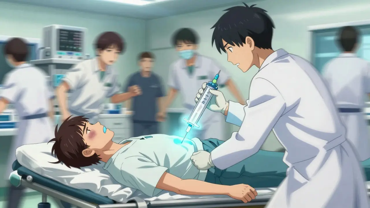
Emergency Care: Immediate Actions and Hospital Treatment
If you or someone else experiences sudden chest pain and difficulty breathing, call emergency services immediately. Do not wait to see if it passes. While waiting for help, keep the person calm and sitting upright. Lying flat can make breathing harder. Loosen any tight clothing around the chest and neck.
Upon arrival at the hospital, the focus is on stabilization and diagnosis. For suspected tension pneumothorax, doctors will not wait for X-rays. They will perform needle decompression-inserting a large needle into the chest cavity to release trapped air-within minutes. This buys time for definitive treatment.
For stable patients, diagnosis typically involves a chest X-ray, which detects air in the pleural space with high accuracy. However, newer protocols increasingly use point-of-care ultrasound (E-FAST), which is faster and highly sensitive. Once diagnosed, treatment depends on the size of the collapse and the patient's symptoms:
- Observation: Small collapses (<15-30%) in stable patients may resolve on their own. Doctors often administer supplemental oxygen, which speeds up the reabsorption of air from the pleural space.
- Needle Aspiration: A needle is inserted to suck out the air. This works well for primary spontaneous cases but has a moderate success rate.
- Chest Tube Insertion: For larger collapses or secondary pneumothorax, a tube is inserted between the ribs and connected to a suction device to remove air and fluid. This is the standard for most significant cases.
- Surgery (VATS): Video-assisted thoracoscopic surgery is used for recurrent cases or persistent air leaks. Surgeons remove the blebs causing the leak and may roughen the pleural surface (pleurodesis) to prevent future collapses.
| Treatment Method | Best For | Success Rate / Notes |
|---|---|---|
| Observation + Oxygen | Small primary collapses (<2cm rim) | 82% resolve spontaneously within 14 days |
| Needle Aspiration | Larger primary spontaneous cases | 65% immediate success; less invasive than tubes |
| Chest Tube (Thoracostomy) | Secondary pneumothorax, large collapses, trauma | 92% success; requires hospital stay; risk of infection |
| VATS Surgery | Recurrent cases, bilateral issues, persistent leaks | 95% long-term success; reduces recurrence to 3-5% |

Who Is at Risk? Understanding Causes and Triggers
Not everyone is equally likely to suffer a collapsed lung. Primary spontaneous pneumothorax disproportionately affects tall, thin males aged 20-40. Why? The mechanics of lung growth in tall individuals create higher stress on the apical (top) parts of the lungs, leading to the formation of fragile air blisters called blebs. When these burst, air escapes.
Smoking is the single biggest modifiable risk factor. Smokers have a significantly higher incidence of pneumothorax compared to non-smokers. In fact, quitting smoking reduces the risk of recurrence by nearly 77% within a year. Other risk factors include:
- Underlying Lung Disease: Conditions like Chronic Obstructive Pulmonary Disease (COPD), pneumonia, or tuberculosis weaken lung tissue.
- Family History: Genetics can play a role, particularly in connective tissue disorders like Marfan syndrome.
- Recent Medical Procedures: Any procedure involving the chest or upper abdomen carries a small risk of accidental puncture.
- Pressure Changes: Scuba diving or flying shortly after a previous episode can trigger a recurrence due to changes in atmospheric pressure.
Recovery and Preventing Recurrence
Recovering from a pneumothorax takes patience. Even after the lung re-expands, the tissue needs time to heal. Most patients can return to light activities within a few weeks, but full recovery may take months. Follow-up chest X-rays are crucial to ensure the lung stays expanded and to check for any delayed complications.
Recurrence is a real concern. About 30-50% of people who have one spontaneous pneumothorax will experience another. To minimize this risk:
- Quit Smoking: This is non-negotiable for lung health. Avoid secondhand smoke as well.
- Avoid Pressure Changes: Do not fly or scuba dive until your doctor clears you. Typically, you should wait at least 2-3 weeks after resolution before flying.
- Monitor Symptoms: Know the warning signs. If chest pain returns, seek care immediately.
- Consider Surgery: If you’ve had two episodes, discuss VATS surgery with a thoracic specialist. It dramatically lowers the chance of a third occurrence.
Living with a history of pneumothorax doesn’t mean you’re doomed to repeat it. With proper care and lifestyle adjustments, most people live normal, active lives. The key is vigilance and working closely with your healthcare team.
How quickly does a collapsed lung happen?
A primary spontaneous pneumothorax often happens suddenly, sometimes during sleep or routine activity. Tension pneumothorax can develop within minutes following trauma or a procedure, making it a rapid-onset emergency.
Can a collapsed lung heal on its own?
Yes, small primary pneumothoraces (less than 15-30% collapse) often resolve spontaneously within 1-2 weeks with observation and supplemental oxygen. Larger collapses or those in patients with lung disease usually require intervention like a chest tube.
Is a collapsed lung fatal?
While rare, a tension pneumothorax can be fatal if not treated immediately because it compresses the heart and major vessels. Prompt medical attention drastically reduces mortality rates, especially for primary cases.
When can I fly after a collapsed lung?
Most guidelines recommend waiting at least 2-3 weeks after complete resolution confirmed by X-ray before flying. Always consult your doctor, as individual healing times vary.
Does smoking increase the risk of recurrence?
Yes, significantly. Smoking damages lung tissue and impairs healing. Quitting smoking reduces the risk of recurrent pneumothorax by approximately 77% within one year.