How the Immune System Contributes to Reperfusion Injury
Immune Response Treatment Calculator
Patient Risk Assessment
Treatment Options
Patient Risk Score
Your patient has a high risk of reperfusion injury based on their clinical profile.
Risk factors identified: Prolonged ischemia duration, diabetes, and hypertension.
This risk score aligns with the article's take-home checklist: high-risk patients benefit most from early immunomodulatory therapy.
When a blocked artery finally reopens after a heart attack or stroke, doctors celebrate the restored blood flow. Yet that very rush of oxygen‑rich blood can paradoxically worsen the injury. The hidden culprit? Our own immune system. Below we unpack why the immune response flips from protector to aggressor during reperfusion injury and what that means for treatment.
Key Takeaways
- Reperfusion injury is driven by a burst of oxidative stress and a cascade of immune‑mediated inflammation.
- Both innate cells (like neutrophils) and adaptive components (T‑cells, antibodies) aggravate tissue damage.
- Targeting specific immune pathways-such as the complement cascade or cytokine signaling-shows promise in animal models and early clinical trials.
- Future therapies will likely combine timely reperfusion with precise immunomodulation.
Understanding Reperfusion Injury
Reperfusion Injury is the tissue damage that occurs when blood supply returns to tissue after a period of ischemia, triggering oxidative stress and inflammation. The phenomenon was first described in the 1960s during heart‑bypass surgery, but it is now recognized across organs-heart, brain, kidney, and even skeletal muscle.
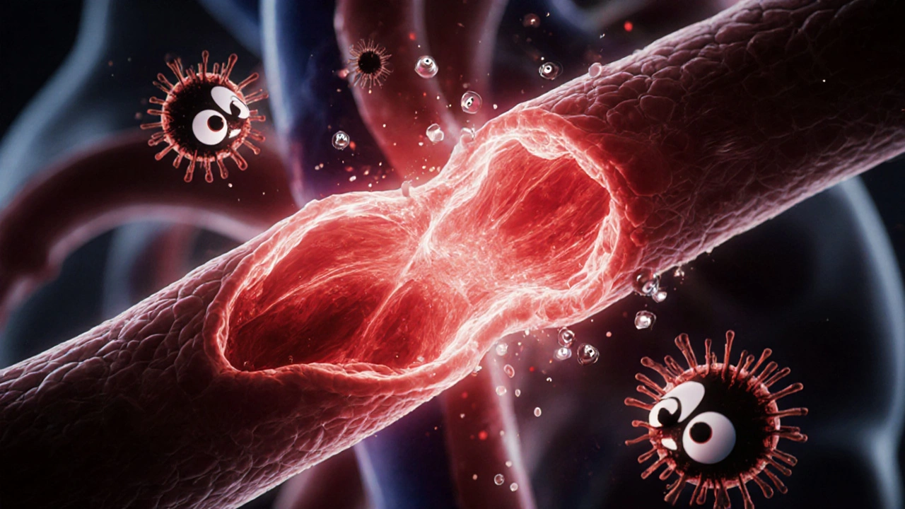
The core events are simple yet lethal: oxygen floods the previously starved cells, generating a surge of reactive oxygen species (ROS). Simultaneously, damaged endothelial cells become sticky, allowing white blood cells to rush in. The result is a perfect storm of cellular death, swelling, and microvascular blockages that can undo the benefits of re‑establishing blood flow.
The Immune System’s Double‑Edged Role
Immune System is the body’s network of cells, proteins, and signaling pathways that defend against infection and help heal injury. In the context of reperfusion, the immune system swings from healing to harming within minutes.
Two major arms are at play:
- Innate immunity-the rapid, non‑specific response involving neutrophils, macrophages, the complement cascade, and pattern‑recognition receptors.
- Adaptive immunity-the slower, antigen‑specific response driven by T‑cells and antibodies.
Both arms release cytokines, chemokines, and ROS that amplify tissue injury.
Innate Immune Cells: First Responders That Overstay Their Welcome
Neutrophils are the most abundant type of white blood cell, acting as the front‑line defenders against infection and injury. Within minutes of reperfusion, they adhere to activated endothelium, transmigrate, and release granular enzymes (myeloperoxidase, elastase) plus massive amounts of ROS. Their extracellular traps (NETs) provide a scaffold for platelet aggregation, worsening microvascular obstruction.
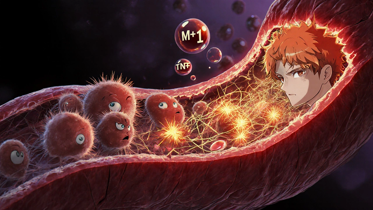
Macrophages, especially the classically activated M1 phenotype, secrete tumor necrosis factor‑α (TNF‑α) and interleukin‑1β (IL‑1β), further recruiting neutrophils and amplifying inflammation. In contrast, the alternatively activated M2 phenotype appears later, attempting to resolve inflammation, but the early M1 surge often sets irreversible damage.
Adaptive Immunity: The Unexpected Player
While traditionally viewed as a slower response, adaptive components can be primed by ischemic antigens released during the first injury wave. T cells are a subset of lymphocytes that recognize specific antigens and orchestrate immune responses infiltrate the injured tissue within hours, producing interferon‑γ (IFN‑γ) that sustains M1 macrophage activation.
Autoantibodies against oxidized phospholipids and heat‑shock proteins have been detected in post‑myocardial infarction patients, suggesting that humoral immunity also contributes to delayed reperfusion injury.
Key Molecular Mediators
Reactive Oxygen Species are highly reactive molecules formed by the incomplete reduction of oxygen, capable of damaging DNA, proteins, and lipids. ROS generation spikes during reperfusion due to mitochondrial electron‑transport chain dysfunction and NADPH oxidase activation in neutrophils.
Cytokines are small proteins that act as messengers between cells during immune responses such as IL‑6, IL‑1β, and TNF‑α create a feedback loop that recruits more immune cells and increases vascular permeability.
The Complement System is a cascade of plasma proteins that opsonize pathogens and promote inflammation. Components C3a and C5a act as chemotactic factors, drawing neutrophils and monocytes into the reperfused zone.
Endothelial Cells line blood vessels and, when activated, express adhesion molecules (ICAM‑1, VCAM‑1) that tether leukocytes. Their dysfunction also leads to a loss of nitric oxide, worsening vasoconstriction.
Therapeutic Strategies Targeting Immunity
Because the immune surge is both rapid and damaging, timing is crucial. Several approaches are under investigation:
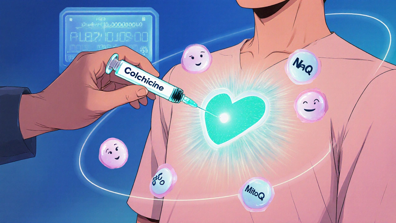
- Antioxidants - High‑dose N‑acetylcysteine (NAC) and mito‑targeted antioxidants (MitoQ) scavenge ROS, but clinical results remain mixed.
- Neutrophil inhibition - Agents like colchicine, which suppress microtubule polymerisation, reduce neutrophil activation and have shown a modest reduction in infarct size in recent trials.
- Complement blockade - C5‑blocking antibodies (e.g., eculizumab) curb the C5a‑driven chemo‑attraction; early-phase studies in myocardial infarction patients report lower troponin peaks.
- Cytokine neutralisation - IL‑1β antagonists (canakinumab) lower subsequent cardiovascular events, hinting at a role in the reperfusion window.
- Regulatory T‑cell (Treg) therapy - Adoptive transfer of Tregs tempers the pro‑inflammatory milieu; animal data show improved perfusion and reduced scar formation.
Importantly, these immunomodulators are most effective when paired with rapid reperfusion techniques (PCI, thrombolysis) and are administered within the first 30‑60 minutes of blood flow restoration.
Future Directions and Research Gaps
While pre‑clinical models have highlighted dozens of immune targets, translating them to bedside care faces hurdles:
- Patient heterogeneity - Age, comorbidities, and genetic background affect immune responsiveness.
- Biomarker timing - Real‑time measurement of cytokine bursts or complement activation could guide individualized therapy.
- Combination therapy - Simultaneously dampening ROS, neutrophil adhesion, and complement may produce synergistic benefits but requires careful safety profiling.
Advances in single‑cell RNA sequencing are already teasing the precise phenotypes of infiltrating immune cells, opening the door to highly specific interventions that spare protective mechanisms while curbing damage.
Take‑Home Checklist for Clinicians and Researchers
- Identify high‑risk patients (e.g., prolonged ischemia, diabetes) where immune‑targeted adjuncts may be most valuable.
- Consider early administration of a single‑target agent (colchicine, NAC) within the first hour of reperfusion.
- Monitor biomarkers such as hs‑CRP, IL‑6, or complement split products to gauge treatment response.
- Stay alert for emerging trial data on complement inhibitors and Treg‑based therapies.
Frequently Asked Questions
What exactly is reperfusion injury?
Reperfusion injury occurs when blood returns to tissue after a period without oxygen, causing a surge of free radicals and an intense inflammatory response that can enlarge the original damage.
Why does the immune system become harmful after blood flow is restored?
The sudden oxygen influx activates neutrophils and complement proteins, which release reactive oxygen species and cytokines. These molecules damage cell membranes, attract more immune cells, and create micro‑vascular blockages.
Can antioxidants prevent reperfusion injury?
Antioxidants can neutralise some ROS, but clinical trials have shown mixed results. The timing of administration (ideally before or immediately after reperfusion) appears critical for any benefit.
What are the most promising immunomodulatory drugs right now?
Early data suggest complement C5 blockers, IL‑1β antibodies, and colchicine (a neutrophil inhibitor) can reduce infarct size when given promptly. Ongoing phase‑III trials will clarify their place in standard care.
Is there a way to predict who will suffer the worst reperfusion injury?
High‑sensitivity CRP, elevated troponin levels, and genetic polymorphisms in antioxidant enzymes (e.g., SOD2) are being studied as predictors. Combining these markers with imaging (MRI edema) may soon guide personalized therapy.
Valerie Vanderghote
October 17, 2025 AT 12:26When the occluded vessel finally yields to the interventionalist's skill, the body does not simply celebrate a return of oxygen, it launches a biochemical cascade that can paradoxically magnify the original insult; the first wave of reactive oxygen species (ROS) is generated within seconds by mitochondria that have been left in a highly reduced state, and these ROS instantly oxidize lipids, proteins, and DNA in the surrounding myocytes. At the same time, endothelial cells that have been deprived of shear stress begin to express adhesion molecules such as ICAM‑1 and VCAM‑1, turning the inner lining of the vessel into a sticky trap for circulating leukocytes. Neutrophils, the most abundant white blood cells, obey chemotactic cues and swarm into the tissue within minutes, where they release myeloperoxidase, elastase, and a torrent of ROS that further degrade the extracellular matrix. Their extracellular traps (NETs) act like fishing nets that capture platelets, creating micro‑vascular obstructions that prevent even the newly restored blood from reaching the deepest zones of the infarct. Meanwhile, resident macrophages polarise towards an M1 phenotype under the influence of TNF‑α and IL‑1β, secreting additional cytokines that recruit more neutrophils in a self‑reinforcing loop. The complement cascade, once thought to be a mere by‑stander, becomes a central amplifier when C3a and C5a attract even more immune cells and increase vascular permeability, allowing plasma proteins to flood the interstitium and exacerbate edema. Adaptive immunity does not stay silent either; T‑cells that have been primed by ischemia‑derived antigens infiltrate the tissue within hours, releasing interferon‑γ that sustains M1 macrophage activation and impairs the later transition to reparative M2 phenotypes. Autoantibodies against oxidised phospholipids have been detected in patients after myocardial infarction, suggesting that humoral mechanisms can prolong the inflammatory milieu well beyond the acute phase. All of these processes culminate in what clinicians recognise as reperfusion injury: a widening of the infarct size, impaired contractile recovery, and a higher likelihood of adverse remodeling. Understanding the precise timing of each immune wave is therefore not an academic exercise but a prerequisite for designing interventions that can be administered within the narrow therapeutic window of the first hour after reperfusion. Researchers are currently testing agents that block specific complement components, inhibit neutrophil adhesion, or scavenge ROS, hoping to blunt the cascade without compromising the essential clean‑up functions of the immune system. The challenge remains to balance dampening the harmful surge while preserving the protective aspects that are necessary for eventual tissue repair. In practice, the most promising strategies appear to be those that combine rapid mechanical reperfusion with a targeted pharmacologic adjunct given immediately at the bedside. As we move forward, the integration of real‑time biomarkers-such as high‑sensitivity CRP, IL‑6, or complement split products-with point‑of‑care imaging may allow clinicians to personalise therapy on a per‑patient basis, tailoring immunomodulation to the individual's inflammatory signature. Ultimately, the goal is to turn the immune system from a double‑edged sword back into a scalpel that precisely cuts away the damaged tissue while sparing the viable myocardium.
Michael Dalrymple
October 23, 2025 AT 07:20In the broader philosophical context, the immune response to reperfusion can be viewed as an embodiment of the dialectic between order and chaos; the rapid restoration of blood flow represents a moment of synthesis, yet the ensuing inflammation reveals the antithetical forces at play. By appreciating this tension, clinicians can adopt a more nuanced approach that does not indiscriminately suppress immunity, but rather modulates specific pathways to restore homeostasis.
Emily (Emma) Majerus
October 29, 2025 AT 01:13Great summary, thx!
Virginia Dominguez Gonzales
November 3, 2025 AT 20:06The dramatic flair of neutrophil infiltration reminds me of a battlefield where the first wave of soldiers, eager yet reckless, storms the gates only to find that their weapons are as much a curse as a boon. Their release of NETs is like a double‑edged sword, ensnaring foes while also trapping the very reinforcements needed for recovery. This paradox is why targeted therapies that quiet the overzealous troops without disbanding the entire army are so vital.
Carissa Padilha
November 9, 2025 AT 15:00Everyone talks about antioxidants like they're the holy grail, but what if the pharma giants are pushing them to keep us dependent on endless supplement cycles? The real issue might be that the complement inhibitors are being held back until the next big patent can be cashed in, leaving patients to suffer the preventable damage of unchecked immune cascades. It's a pattern: introduce a breakthrough, then quietly file for extensions while the public remains none the wiser.
Richard O'Callaghan
November 15, 2025 AT 09:53i cant help but notice that the timing of icometric adjuvants is often misreprented in trials; they claim early admin but the protocol start times are later than the realease of blood flow, which makes the data suspecious at best.
Emily Rankin
November 21, 2025 AT 04:46Imagine a future where single‑cell sequencing maps every infiltrating lymphocyte in real time, granting us the power to silence only the destructive clones while bolstering the reparative ones. Such precision would transform reperfusion therapy from a blunt instrument into a symphony of orchestrated cellular responses, echoing the harmonious balance nature strives for.
Rebecca Mitchell
November 26, 2025 AT 23:40Sounds promising but we need data fast.
Frank Diaz
December 2, 2025 AT 18:33The prevailing narrative glorifies rapid reperfusion without acknowledging the ethical dilemma of exposing patients to experimental immunosuppression; we must question whether the pursuit of smaller infarct sizes justifies the potential long‑term compromise of immune competence. Moreover, the reliance on surrogate markers like troponin peaks can obscure the nuanced outcomes that truly matter to patients' quality of life.
Mary Davies
December 8, 2025 AT 13:26While the literature is awash with hopeful headlines, the reality on the ground is that many clinicians remain hesitant to adopt these adjuncts, fearing off‑label liability and the unknown long‑term sequelae of dampening the immune response. This cautious stance, though understandable, may stall the very progress needed to reduce the burden of reperfusion injury.