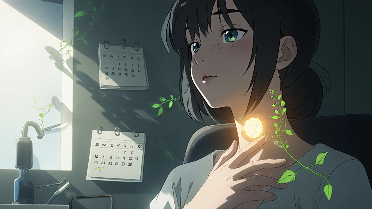
Thyroid Nodules: How to Tell Benign from Cancerous and When a Biopsy Is Truly Needed
Most people with thyroid nodules never know they have them. They show up on an ultrasound done for another reason, or a doctor feels a lump during a routine checkup. The good news? Thyroid nodules are incredibly common-up to 67% of adults over 60 have at least one. The even better news? More than 90% of them are harmless. But that 5-10% that might be cancer? That’s where things get urgent. The key isn’t panic. It’s knowing when to act.
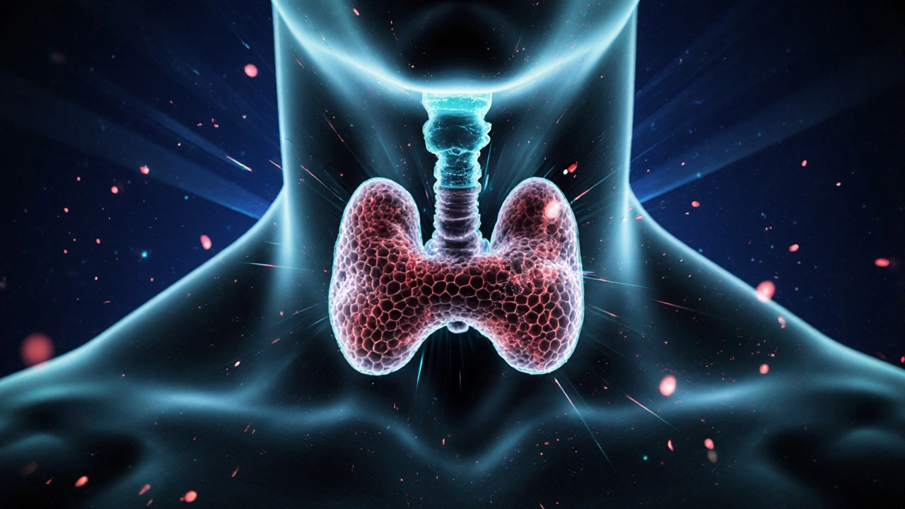
What Makes a Thyroid Nodule Suspicious?
Not all nodules are created equal. Some are just fluid-filled sacs (cysts) or clumps of normal thyroid tissue (colloid nodules). These almost never turn cancerous. Others have features that raise red flags. Ultrasound is the first and most important tool. It doesn’t just show size-it shows structure.Malignant nodules often have specific traits:
- Microcalcifications-tiny white specks that look like sand under the scope. Found in over half of papillary thyroid cancers.
- Irregular or spiculated margins-edges that aren’t smooth, more like jagged lines.
- Hypoechogenicity-appearing darker than the surrounding thyroid tissue. This is one of the strongest predictors.
- Increased blood flow inside the nodule, not just around it.
Benign nodules usually look different: rounded, with smooth edges, maybe partly cystic, or even spongiform-meaning they look like a honeycomb under ultrasound. These are almost always safe.
Size Matters, But Not the Way You Think
You’ve probably heard that nodules over 1 cm need a biopsy. That’s true-but only if they have suspicious features. A 1.5 cm nodule with smooth edges and no calcifications? It’s likely fine. A 7 mm nodule with microcalcifications and irregular borders? That’s the one you worry about.The American Thyroid Association guidelines are clear:
- Biopsy is recommended for nodules ≥1 cm with suspicious ultrasound features.
- For nodules ≥1.5 cm without suspicious features, biopsy is still advised.
- Any nodule ≥2 cm should be biopsied, no matter how it looks.
But size alone isn’t enough. Growth rate is becoming just as important. A nodule that grows more than 2 mm per year in at least two dimensions has a significantly higher chance of being cancerous. This isn’t theoretical-studies show a 3.2 times higher risk for nodules growing faster than this. If your nodule grew from 1.2 cm to 1.6 cm in 12 months? That’s 4 mm of growth. That’s a red flag.
The Bethesda System: Decoding the Biopsy Result
If a biopsy is done, the result is classified using the Bethesda System. It’s not just “cancer” or “not cancer.” It’s a six-tiered scale that tells you the risk level:- Category 1: Nondiagnostic-too few cells. Need a repeat biopsy. Risk: 1-4%
- Category 2: Benign-normal cells. Risk: 0-3%. Usually just monitor.
- Category 3: Atypia of undetermined significance-cells are weird, but not clearly cancerous. Risk: 5-15%.
- Category 4: Follicular neoplasm-cells suggest a tumor, but can’t tell if it’s benign or malignant without surgery. Risk: 15-30%.
- Category 5: Suspicious for malignancy-strong signs of cancer. Risk: 60-75%.
- Category 6: Malignant-cancer confirmed. Risk: 97-99%.
Categories 3 and 4 are the gray zone. That’s where molecular testing comes in. Tests like Afirma GSC or ThyroSeq v3 look at the DNA and RNA inside the cells. They can tell you if a “suspicious” nodule is actually low-risk. One study showed these tests cut unnecessary surgeries by 35% in indeterminate cases. That’s thousands of people avoiding surgery they didn’t need.

What Kind of Cancer Are We Talking About?
If it’s cancer, it’s almost certainly papillary thyroid carcinoma-about 80% of cases. This type grows slowly, often spreads to lymph nodes in the neck, but has a near 100% survival rate when caught early. Follicular carcinoma makes up 10-15%. It rarely spreads to lymph nodes but can travel through the blood to lungs or bones. That’s why it’s harder to catch early.Other types-medullary, anaplastic, lymphoma-are rare. Anaplastic is aggressive and fast-growing, but it’s extremely uncommon. Most thyroid cancers aren’t the kind that kill quickly. They’re the kind that, if left alone, might grow slowly for years before causing trouble.
When Do You Actually Need a Biopsy?
Let’s cut through the noise. Here’s when you need a biopsy:- Your nodule is 1 cm or larger AND has one or more suspicious ultrasound features (microcalcifications, irregular edges, hypoechogenicity).
- Your nodule is 1.5 cm or larger-even if it looks fine on ultrasound.
- Your nodule is 2 cm or larger, period.
- Your nodule has grown more than 2 mm per year in two dimensions, even if it’s under 1 cm.
- You have symptoms like trouble swallowing, hoarseness, or a persistent lump feeling-even if the nodule is small.
And here’s when you probably don’t:
- Your nodule is under 1 cm, looks perfectly benign on ultrasound, and hasn’t grown in two years.
- You’re over 70 with a small, stable nodule and no family history of thyroid cancer.
- You have multiple tiny nodules (common in Hashimoto’s) and no suspicious features.
Overdiagnosis is real. Since the 1970s, thyroid cancer diagnoses have increased 15-fold-not because more people are getting sick, but because we’re scanning more and finding tiny, harmless nodules. Many of these would never cause harm in a person’s lifetime. Biopsying every single nodule leads to unnecessary surgeries, scarring, and lifelong hormone replacement.
What Happens After the Biopsy?
If the result is benign (Category 2), you’ll likely be monitored with ultrasound every 12-24 months. If it’s indeterminate (Categories 3 or 4), molecular testing is the next step. If it’s positive (Categories 5 or 6), you’ll be referred to a thyroid surgeon. Most of the time, only half the thyroid is removed (a lobectomy). If cancer is confirmed, you’ll need radioactive iodine treatment and lifelong thyroid hormone replacement.But here’s the new frontier: active surveillance. For very small papillary cancers (under 1 cm) with no spread, some patients are now being monitored with yearly ultrasounds instead of surgery. A 2021 study showed 87% of these tiny cancers didn’t grow over five years. For low-risk patients, watching and waiting is now a valid option.
What You Can Do Right Now
If you’ve been told you have a thyroid nodule:- Ask for the ultrasound report. Don’t just take the doctor’s word-get the details. Look for size, shape, calcifications, margins.
- Ask if growth has been tracked. If you’ve had an ultrasound before, compare measurements. Did it grow more than 2 mm in a year?
- Ask about molecular testing if your biopsy is indeterminate. It can save you from surgery.
- Don’t rush into surgery for a small, stable nodule without a second opinion.
- Find a specialist who follows the American Thyroid Association guidelines. Not all doctors do.
Thyroid nodules aren’t a crisis. They’re a puzzle. And the pieces-size, shape, growth, and biopsy results-tell the story. You don’t need to fear every lump. You just need to know when to listen.
Cris Ceceris
November 11, 2025 AT 02:29Man, I had no idea thyroid nodules were this common. My grandma had one and they just watched it for years-turns out she was lucky. I always thought any lump = cancer, but this breaks it down so clearly. The part about growth rate being more important than size? That’s wild. I’m gonna start tracking my own ultrasounds now.
Erika Puhan
November 12, 2025 AT 15:07While the author cites ATA guidelines, they conspicuously omit the fact that overdiagnosis leads to iatrogenic harm-particularly in elderly populations where low-risk papillary carcinoma often remains clinically silent. The normalization of molecular testing as a panacea ignores cost-benefit asymmetries in resource-constrained systems. Furthermore, the emphasis on ultrasound morphology lacks validation in heterogeneous populations, particularly those with iodine deficiency.
Edward Weaver
November 13, 2025 AT 07:03Ugh, another liberal ‘don’t panic’ article. If you’re American and you’ve got a nodule, you get scanned, tested, and treated. Period. Other countries wait and see until it’s too late. We’re not playing Russian roulette with cancer here. If the ATA says biopsy at 1.5cm, then biopsy at 1.5cm. No ‘maybe’-this isn’t yoga class.
Lexi Brinkley
November 13, 2025 AT 23:34Okay but like… microcalcifications?? 🤯 I just got my ultrasound results and they said ‘suspicious features’ and I thought I was gonna die. But now I’m like… maybe not?? 😅 Also, active surveillance?? That’s a thing?? I’m gonna print this out and show my doc. 🙏
Kelsey Veg
November 14, 2025 AT 20:16so like… if your nodule is under 1cm and no calcifcations and no growth… you just chill? no biopsy? but what if it’s cancer and you miss it?? like… i dont trust doctors anymore, they’re all in it for the money. i read this one guy on reddit who said his nodule turned into cancer in 6 months and he didn’t even know. 😳
Alex Harrison
November 16, 2025 AT 01:09Good breakdown. I had a Category 3 result last year and my doc didn’t mention molecular testing until I asked. Turns out my test came back low risk-saved me from a lobectomy. Seriously, if you get an indeterminate result, push for Afirma or ThyroSeq. It’s not expensive and insurance usually covers it. Don’t just take ‘watch and wait’ as the only option.
Jay Wallace
November 16, 2025 AT 21:23Let me be clear: the American Thyroid Association guidelines are the gold standard-no other country’s protocols even come close. The fact that you’re even considering surveillance for a nodule over 1 cm is borderline negligent. In Europe, they wait too long-look at the mortality rates. We have the technology, the data, the guidelines. Use them. Don’t gamble with your life because some blogger said ‘it might be fine.’
Alyssa Fisher
November 18, 2025 AT 12:57What struck me most isn’t the medical details-it’s the cultural shift. We’ve gone from ‘if you feel something, cut it out’ to ‘if it’s slow, quiet, and harmless, let it be.’ That’s not just medicine. That’s wisdom. We used to treat every anomaly as an enemy. Now we’re learning to distinguish between threats and ghosts. The real victory isn’t removing a nodule-it’s choosing not to remove it when you don’t have to. That takes courage, not just science.